The Biochemistry & Biophysics of Cataract Formation
Cataracts progress in stages, but the development of the condition depends on age, exposure to UV experienced over a lifetime, genetic factors and some lifestyle factors, such as smoking, high alcohol consumption or nutritional deficiencies.
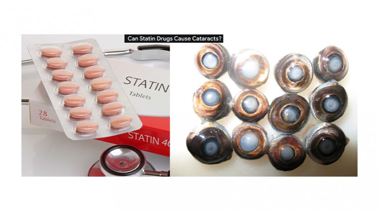
People with diabetes are at higher risk, as are those who take certain prescription medicines, such as corticosteroids or phenothiazine-related medications.
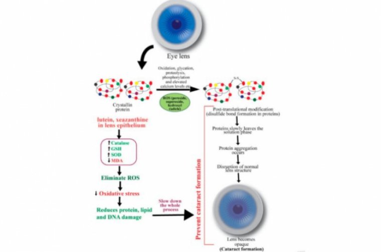
The lens is made up of cells packed with structural proteins called crystallins. Crystallins within each lens cell form a protein-dense gel, and the gel’s optical properties — like its transparency and the way it refracts light — help focus light onto the retina.
Crystallins
Crystallins are the collection of structural proteins found in the lens of the eye that help to focus light onto the retina. We know that over our lifetimes they can accumulate damage, losing their native structure and sticking together to form aggregates – one of several mechanisms that causes cataracts.
According to the Harvard study, the crucial discovery that wild-type (undamaged) crystallin promotes aggregation of mutant (damaged) versions – without itself aggregating. Chemical bonds between sulfur atoms within the protein (disulfide bonds) are found to play a role in aggregation.
The crystallin protein molecules engages in oxidation–reduction reactions with one another – disproving the long-held assumption that crystallins are inert.
Analyzing Crystallin Proteins
In ligth of the analysis, the eye lens proteome is fascinating because it has evolved to minimize light scattering. That means it must resist aggregation because protein aggregates (clumps of many molecules of a protein) scatter visible light.
Yet, the protein molecules in the core region of the lens are synthesized before birth and never replaced thereafter. These protein molecules are undergoing an aging process even before we are born, and they continue to accumulate various kinds of damage throughout life.
The biochemistry and biophysics of this highly concentrated solution of highly aged proteins, and how they have evolved to resist aggregation, has been of huge interest to many researchers over the years.
Of course, as fascinating and unique as crystallins are for their own sake, they also have important implications for public health.
Their aggregation – when it does eventually occur – causes cataracts, which affect tens of millions of patients. Some people falsely assume that modern surgery has solved the problem of cataracts.
In fact, millions of patients around the world never benefit from the surgery, either because they can’t afford it, because they live in areas where it is not available, or because surgery is contraindicated for them.
Moreover, the total cost of cataract surgeries performed worldwide already adds up to tens of billions of dollars per year, and as the global population ages, cataract prevalence will rise dramatically.
The Role of Crystallins in Causing Cataracts
Proteins from the crystallin family make up the lion’s share of all protein molecules in the cells of the eye lens. Since they are never replaced, at least in the lens core region, they accumulate damage over a lifetime.
Eventually these crystallin protein molecules begin to lose their native structure (the normal 3D arrangement of atoms) and stick together to form aggregates.
Once the aggregates reach a size that is comparable to the wavelength of visible light, they begin to scatter light, resulting in less light reaching the retina and blurring of the resulting image.
Because blue light has the shortest wavelengths, it gets scattered the most, so the colors we see also change, becoming yellower.
Mixing Mutated Proteins & Lens Crystallins
During the study the initial observation was striking: mixing mutated protein with normal, unmutated protein led to rapid aggregation and a spike in light scattering.
We were able to use gel electrophoresis, and more recently mass spectrometry, to separate the components of the aggregates and saw, to our surprise, that only the mutant protein was present there.
There are several health conditions elsewhere in the body in which misfolded mutant proteins cause otherwise normal (wild-type) proteins to misfold likewise – the mutant protein acts as a template.
This is the mechanism behind prion diseases (such as Creutzfeldt-Jakob disease), and it leads to aggregation of both the mutant and the wild-type molecules.
The initial hypothesis was that a similar phenomenon could exist in eye lens crystallins. However, the truth turned out to be the reverse: a wild-type crystallin promoted aggregation of a mutant version of itself, without itself aggregating.
This new phenomenon was intriguing, but its mechanism remained totally mysterious. The search for this mechanism led to another surprise: a chemical reaction was taking place between these crystallin protein molecules, a process of oxidation–reduction.
Also it is possible that there is a second aggregation-promoting mechanism at work.
The crystallin proteins can pass disulfide bonds among themselves, from one molecule to another to another. These disulfide bonds are formed when two atoms of sulfur within one protein molecule react with each other. This chemical reaction releases electrons, making it an oxidation reaction.
The disulfide bond can then be transferred to another pair of sulfur atoms on a second molecule of this protein. In chemical terms, molecule 2 releases electrons that are received by molecule 1, so molecule 2 is oxidized and molecule 1 is reduced.
These transfers of disulfides can be passed back and forth for a long time if the two molecules are equivalent.
The Process of Aggregation
There are reactive sulfur atoms in each molecule of gamma-crystallin. In wild-type protein, most of those sulfur atoms are hidden and therefore not available for this kind of chemical reaction.
The situation changes if a mutation or another form of damage causes the protein’s structure to “loosen up”, exposing more sulfur atoms.
If a pair of sulfur atoms that is normally hidden becomes exposed and forms a disulfide bond, the protein becomes trapped in an aberrant structure and becomes sticky, leading to aggregation. Studies suggest that this type of chemistry underlies gamma-crystallin aggregation in the lenses of cataract patients.
Here are the dots. Disulfide bonds are passed around among crystallin molecules like a hot potato: if they land on a damaged protein molecule that, due to its looser structure, displays sulfur atoms that it should have kept hidden, then this damaged molecule gets trapped in a sticky non-native structure, and forced to aggregate.
The disulfides do no great harm to the structurally sound crystallin molecules, and may even be protective, but they drive the structurally weakened molecules into aggregates that scatter light – hence, the aggregates in our experiment only contained mutant (damaged) proteins.
Is It Possible To Stop Proteins From Aggregating?
The cells of the core region of the eye lens cannot make new protein molecules, nor can they actively degrade them. Peptide-based drugs would be expected to be rapidly broken down and metabolized in any other part of the body, but not in the nucleus of the lens.
Although we haven’t yet reported any results with potential peptide drug candidates, we are pursuing several that we believe could inhibit aggregation by affecting both structure and chemistry.
We anticipate that the main challenge will not be finding peptides that can inhibit aggregation, but rather delivering such peptides to the most vulnerable cells of the lens.
Peptide drugs tend to be large molecules, and we don’t yet know if they will penetrate the tissue in sufficient quantities.
Efforts to treat cataracts therapeutically have grown in number and made waves in recent years; at least two distinct lipid-based treatment approaches are now being pursued, for example.
No drugs have been approved so far, and ultimately, a combination of drugs might be needed. But the more we learn about the biochemistry and biophysics of cataract formation, the wider the space of therapeutic possibilities will be.