3D Visualization in Cataract Surgery
The technological advances made in recent years have led to an increase in the number of microsurgeries performed across different surgical specialties.
To overcome the potential limitations of conventional microsurgery using the binocular microscope, development of three-dimensional (3D) display systems and their translational use in medicine has been ground-breaking in the field of microsurgery and in ophthalmic surgery in particular.
Increasingly, more experience with the conventional 3D heads-up display system among both anterior segment and vitreoretinal surgeons is being reported in scientific meetings and high-impact ophthalmology journals.
The use of the active 3D head-mounted display systems in both anterior and posterior segment surgeries is also being increasingly reported. This article is a literature review of the applications of 3D display systems in ophthalmic surgery.
The benefits of 3D visualization in cataract surgery include improved ergonomics, high magnification optics, digital guidance systems that can be implemented in association with it, as well as collaboration with colleagues and educating residents and fellows.
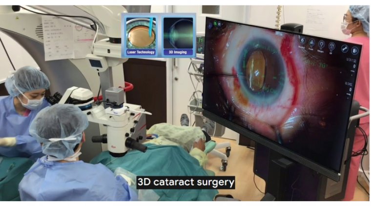
Three-dimensional (3D) digital visualization is a newly developed technology in the domain of eye microsurgery. It consists of one 1080 p camera, central processing unit (CPU), monitor, and polarized glasses.
The camera is wired to the CPU, which processes the live image and displays overlapping stereo images on the widescreen HD monitor. Three-dimensionality is then experienced by donning the circularly polarized glasses.
Advantages of the system involve higher depth of field and working distance at the retinal surface in high magnification, improved field of view, higher dynamic range that reduces instrument glare and illuminates shadows, and digital image filtering to enhance visualization without the use of vital dyes.
While 3D Surgery has become a staple in retinal procedures, it has yet to become prevalent in anterior segment surgery. This may change as surgeons become aware of the benefits a 3D system offers.
According to Ashley R. Brissette, MD, MSc, FRCSC, the degree of visualization through the surgical microscope is quite good, which could prove to be beneficial for physicians as they perform challenging procedures.
“I think that there are other benefits to 3D surgery that will become readily apparent over time,” said Dr. Brissette, assistant professor of ophthalmology, Weill Corneal Medicine and New York Presbyterian Hospital, New York.
A number of factors come into play in the quest for ideal intraoperative visualization when using a surgical microscope.
Dr. Brissette pointed out that during cataract surgery, the microscope must be moved to obtain a better view.
Some structures are always out of focus, the video and recordings can come in and out of focus because of accommodation especially by trainees, and the illumination and magnification must be optimal.
Ergonomics are important to Dr. Brissette. The careers of many ophthalmologists are truncated by back and neck pain caused by extended periods of surgery and examinations over the years.
3D TECHNOLOGY
Technology is currently available (NGenuity, Alcon) (Artveo 300, Zeiss) that provides 4K 3D visualization using 3D glasses. Alcon’s system uses a digital system that processes images and then displays them.
The Zeiss system is one of integrated digital optics, featuring intraoperative optical coherence tomography for corneal surgery and will be introducing digital overlays for toric alignment in cataract surgery, Dr. Brissette explained.
Dr. Brissette noted that the new technology offers exciting options for ophthalmologists. “The three unique advantages of 3D technology are visualization, ergonomics, and education,” she said. “The last has become especially important in anterior segment surgery using the 3D heads-up systems.”
The improvements in visualization seen with 3D technology result from the increased quality of vision to the total operating space and offers high-definition resolution intraoperatively from the nasal to the lateral canthi.
This eliminates looking into the microscope and the frequent adjustments needed to obtain clear focus; the entire field is in focus, she emphasized. Dr. Brissette provided an example of how well 3D technology makes surgery easier.
In a patient who is a high myope undergoing cataract surgery the anterior chamber is deep and using a microscope adjusting the focus is needed to see the nuclear fragments.
Because of 3D technology, there is no need to adjust the focus intraoperatively to visualize the angle during microinvasive glaucoma procedures and for corneal and conjunctival procedures.
Using 3D systems, surgeons can sit back in the chair and maintain a comfortable position during longer cases and days in the operating room. “This feature will help to maintain career longevity,” she said. Education likely is one of the most important benefits of 3D systems.
In complicated cases, personnel from different ophthalmic disciplines as well as residents and fellows can view the surgery simultaneously. Nurses viewing the procedures can anticipate the needs of the surgeons intraoperatively.
No refocusing of the field of view is needed as the various surgeons move in and out of the primary position or for teaching opportunities. Surgical skills can be improved by reviewing cases in 3D with residents and fellows.
“The educational component is becoming more and more important with the education of the next generation of ophthalmologists,” Dr. Brissette noted.
Let's Check What Others Think & Say